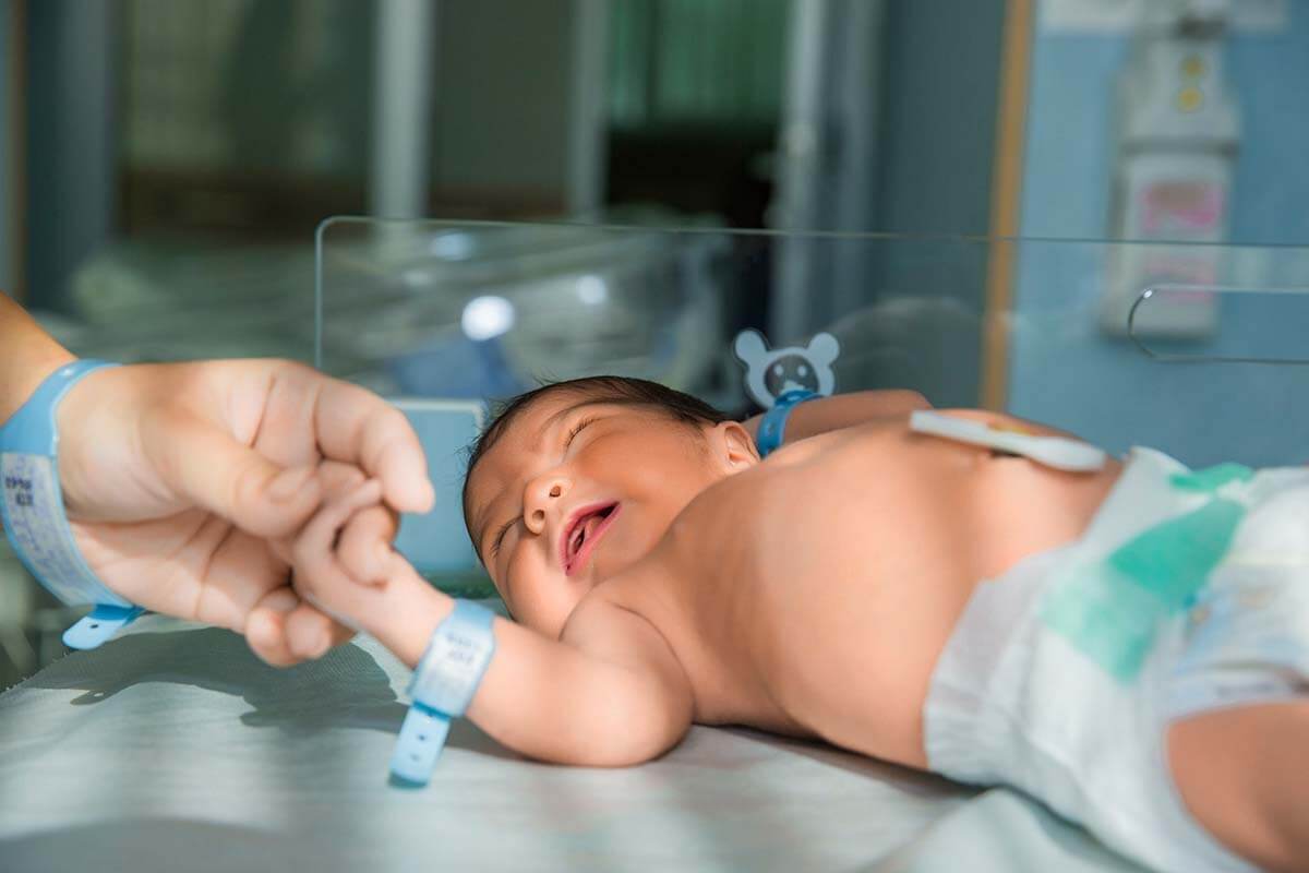
Retinopathy of Prematurity (ROP) is a significant eye health concern commonly observed in premature infants with lower birth weights compared to their full-term counterparts. This condition manifests in the avascular zones of the retina in premature babies, leading to nerve damage and subsequent vision loss.
Notably, Retinopathy of Prematurity often develops without evident symptoms recognizable by parents, underscoring the importance of proactive medical monitoring. Routine check-ups play a pivotal role in the early detection of ROP, facilitating timely intervention and treatment.
This eye disorder poses a particular risk in premature infants, given the underdeveloped nature of their retinas. Left untreated, ROP can progress, resulting in severe nerve damage and permanent vision impairment. Therefore, regular and comprehensive eye examinations are crucial for identifying subtle signs of ROP and initiating prompt therapeutic measures.

What is Retinopathy of Prematurity?
Retinopathy of Prematurity (ROP) is a condition that primarily affects babies born prematurely, specifically those born before 32 weeks of gestation or weighing less than 1500 grams. In these infants, the retinal vessels, crucial for normal vision development, are incompletely formed.
As these underdeveloped retinal vessels continue to mature after birth, they may deviate from their normal course, advancing towards the vitreous gel in front of the retina. This abnormal progression can dislodge the retina, leading to tears and bleeding within the retina. If left unaddressed, this aberrant development of veins can result in severe complications, potentially culminating in total blindness.
Navigating the Stages of Retinopathy of Prematurity:
In understanding the progression of retinopathy of prematurity (ROP), three key concepts come into play: zone, stage, and plus disease.
Zone: The classification spans from Zone 1 to Zone 3, indicating the extent of retinal coverage by intact vessels. Zone 1, characterized by minimal intact vessels, transitions to Zone 3, where very little empty space devoid of intact vessels exists. The associated risk diminishes as one moves from Zone 1 to Zone 3.
Plus Disease: Manifesting as increased tortuosity and dilation in the vessels, plus disease is classified as either present or absent.
Stage: The advancing stages correlate with an increased risk of vision loss attributed to ROP.
- Stage 0: Retinal vessels are underdeveloped, but no other stage findings are present.
- Stage 1: A demarcation line forms at the border between areas with and without retinal vessels.
- Stage 2: The demarcation line thickens, now referred to as a ridge.
- Stage 3: Damaged retinal vessels emerge, moving upward from the retina’s surface toward the vitreous.
- Stage 4: A portion of the retina separates from the underlying layer, indicative of partial retinal detachment.
- Stage 5: The entire retina undergoes separation from the underlying layer, resulting in total retinal detachment.
This structured delineation of zones, stages, and plus disease aids in the comprehensive assessment and management of retinopathy of prematurity, guiding healthcare professionals in tailoring interventions to mitigate the risk of vision impairment in affected infants.
Understanding the Causes and Retinopathy of Prematurity
Retinopathy of Prematurity (ROP) primarily arises due to premature birth, with infants born before 32 weeks (and occasionally below 34 weeks) or weighing less than 1500 grams being particularly susceptible. The critical factor in the development of ROP is the premature nature of the birth, underscoring the importance of gestational age and birth weight as key contributors to this eye condition.
Infants at Risk for Premature Retinopathy Development
The likelihood of premature retinopathy increases as gestational age and birth weight dec
rease. Factors amplifying this risk include compromised lung development, infants requiring ventilator support, a history of infections, cerebral hemorrhage, low levels of insulin-like growth factor 1 (IGF-1), elevated blood sugar, and increased protein excretion in the urine. These multiple risk factors collectively heighten the susceptibility of certain infants to the development of Retinopathy of Prematurity (ROP).
How does Retinopathy of Prematurity Occur?
The occurrence of Retinopathy of Prematurity (ROP) is intricately linked to the premature birth of infants, where retinal vessels haven’t fully developed. At birth, there is an abrupt cessation of vascular development, and this suppression persists, particularly in cases where infants require prolonged respiratory support on ventilators. A crucial factor in this process is VEGF (vascular endothelial growth factor), a substance the body produces to facilitate ongoing vascular development. When VEGF levels become excessively elevated, it triggers abnormal vascular growth, leading to the onset of premature retinopathy. In essence, the disruption of normal vascular development, compounded by extended respiratory assistance and imbalances in VEGF, contributes to the development of ROP in premature infants.
What are the symptoms of Retinopathy of Prematurity?
Detecting Retinopathy of Prematurity (ROP) in its early stages is challenging as there are no discernible symptoms noticeable to families. Consequently, in our country, it is standard practice to screen all infants born at 34 weeks or weighing less than 1500 grams for ROP. The key responsibility for families lies in ensuring timely attendance to all recommended check-ups scheduled by their healthcare providers.
Left untreated, ROP can manifest symptoms in the later stages, including eye deviation (strabismus), absence of the red eye reflex, a white reflex emanating from the eye, and the infant’s failure to track objects with their eyes. It is crucial to emphasize that untreated retinopathy of prematurity carries the potential for complete blindness. Hence, vigilant adherence to medical appointments and early intervention are paramount in preserving the visual health of premature infants.
Diagnosis of Retinopathy of Prematurity
Premature retinopathy is diagnosed through meticulous examination, primarily utilizing an eyedropper and ROP scans conducted with a specialized examination device known as indirect ophthalmoscopy. The optimal timing for ROP screenings is elaborated upon in subsequent sections of our article.
Treatment Methods for Retinopathy of Prematurity
The treatment options for retinopathy of prematurity include intraocular anti-VEGF injections, diode laser therapy, and, if required, vitrectomy surgery. The choice of treatment is determined based on the specific zone and stage of the disease.
Anti-VEGF Injection Therapy:
The administration of intraocular anti-VEGF injections for retinopathy of prematurity does not necessitate general anesthesia. This procedure is conducted under neonatal intensive care conditions or in a sterile environment, utilizing topical anesthesia drops. Specially designed tiny needles for infants are employed for the injection. Following the procedure, the expectation is that abnormal vessels will regress, paving the way for the development of new, healthy vessels. In cases where intraocular injection proves insufficient or the disease reactivates, diode laser application or vitrectomy surgery may become necessary.
Diode Laser Photocoagulation Application
Diode laser therapy for retinopathy of prematurity is undertaken under general anesthesia. Laser pulses are applied to all non-vascularized areas of the retina during this procedure, inhibiting subsequent vascular development in these areas.
Vitreoretinal Surgery (Vitrectomy)
Vitrectomy surgery for premature retinopathy closely mirrors the approach used in adults.
Advancements in Retinopathy of Prematurity Treatment:
In recent years, the treatment landscape for retinopathy of prematurity has witnessed notable progress, particularly with the introduction of intraocular anti-VEGF injections. The success rates have seen a marked increase, as evidenced by findings from the BEATROP study. Notably, the study revealed a significantly lower recurrence rate of 4.3% with anti-VEGF treatment compared to 21.9% with diode laser treatment in cases of stage 3 disease. This statistically significant difference underscores the efficacy and promising outcomes associated with the use of intraocular anti-VEGF injections in advancing the treatment of retinopathy of prematurity
Frequently Asked Questions about Retinopathy of Prematurity:
- What is the First Examination Time for Retinopathy of Prematurity?
According to international guidelines, screening for retinopathy of prematurity begins four weeks after birth for babies born at 27 weeks or later. For infants born at or before 26 weeks, the initial examination occurs when the baby reaches the 31st week.
- How is ROP Examination Done in Babies?
Prior to the examination, eye drops are administered to dilate the pupils. After dilation, a numbing drop is applied. The examination involves indirect ophthalmoscopy using a hand-held lens and a depressor to gently press on the eye.
- What are the reasons for bloodshot eyes in babies after ROP examination?
Eye redness post-examination is common and often attributed to the use of a depressor during the exam. While it may cause initial concern, this bleeding typically heals spontaneously within 10-15 days and does not necessitate treatment. If redness persists with flaking, consulting an ophthalmologist is recommended.
- What Problems May Babies with ROP Encounter in the Future?
Even with treatment, babies with retinopathy of prematurity are at a higher risk of requiring glasses in the future, along with potential issues like strabismus, amblyopia, and retinal tears.
- Situations That May Occur in the Future in Untreated Babies with Retinopathy of Prematurity:
Immediate treatment is crucial for babies diagnosed with retinopathy of prematurity. Left untreated, there is a risk of complete blindness.
- What are the Rates of Retinopathy of Prematurity in Babies?
Annually, approximately 14,000 premature babies in the United States are diagnosed with ROP. Of these cases, 8% to 11% require treatment. According to CRYO-ROP study data, ROP develops in 66% of babies with a birth weight below 1,250 g and in 82% of babies with a birth weight below 1,000 g.

Causes and Treatment of Eye Floaters
Occasionally, individuals may notice floating objects in their field of vision. These can take various [...]
Devamını OkuyunMay

What is Vitreous? How is Vitreous Dysfunction Treated?
Vitreous, derived from the Latin term meaning “glassy,” is a clear gel found between the [...]
Devamını OkuyunMay

Vision Wellness Essentials: Simple and Effective Tips for Eye Health
Our eyes, vital and health-sensitive organs, demand special care to combat conditions such as eye [...]
Devamını OkuyunJan

Nourishing Your Eyes: A Guide to Beneficial Foods for Optimal Eye Health
Caring for our eyes extends beyond weight control; it encompasses exercise, abstinence from smoking and [...]
Devamını OkuyunJan

Understanding Glaucoma: Unveiling the Silent Threat to Vision
Glaucoma, also colloquially known as “black water,” refers to elevated eye pressure. This insidious eye [...]
Devamını OkuyunJan

What is Bietti Crystalline Dystrophy? Can Bietti Crystalline Dystrophy Be Treated?
Bietti Crystalline Dystrophy, identified by Italian physician Dr. Lucas Bietti in 1937, is a rare [...]
Devamını OkuyunJan

Understanding Color Blindness: Symptoms and Treatment
Color blindness is a vision condition where individuals struggle to distinguish specific colors or shades. [...]
Devamını OkuyunDec

What is Uveitis? What are Types, Symptoms, and Treatment Methods?
Uveitis, a severe eye condition characterized by inflammation, is also known as iridocyclitis. Accurate diagnosis [...]
Devamını OkuyunDec