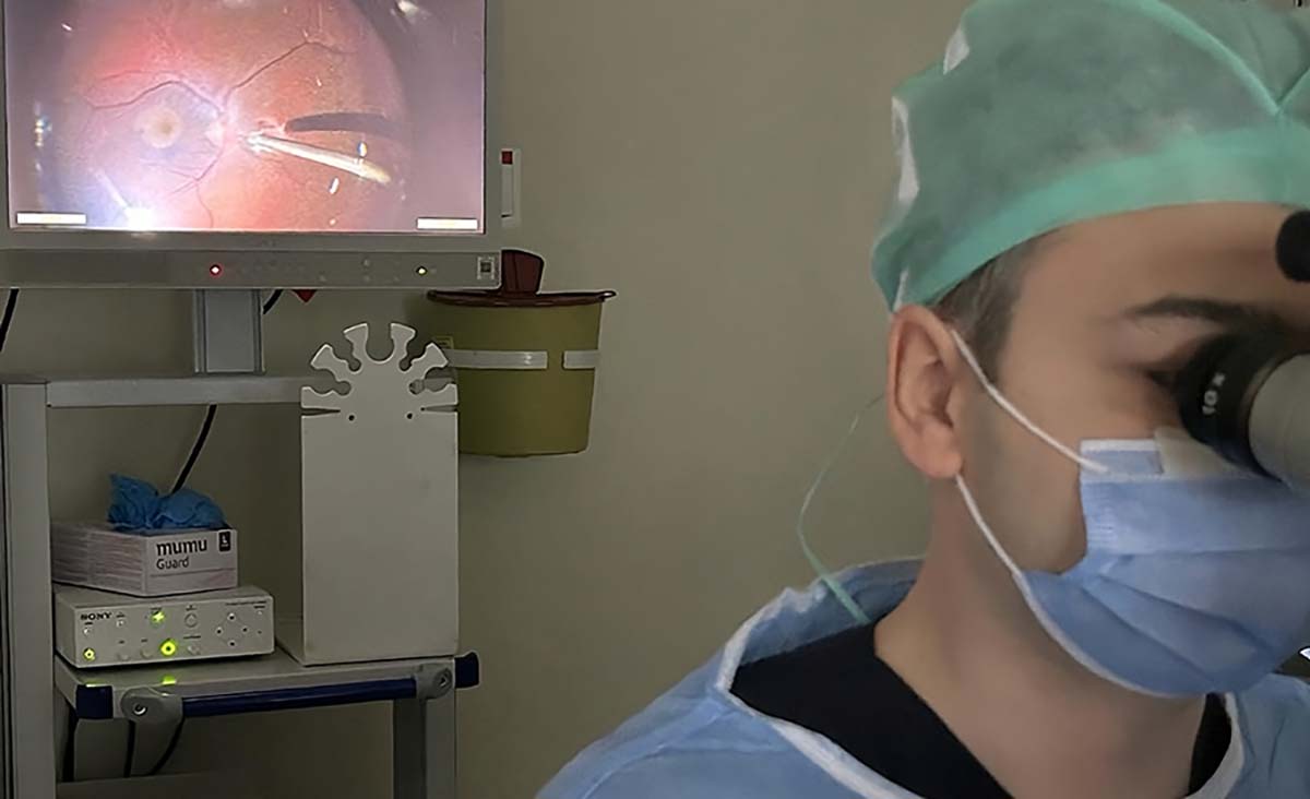
Retinal detachment is a severe eye condition that can lead to profound vision loss or even blindness. Various types of retinal detachment exist, but the most prevalent form involves a tear in the retina. In this article, we will explore rhegmatogenous reti nal detachment, shedding light on its definition, symptoms, diagnostic procedures, and treatment approaches.

What is Retinal Detachment?
Retinal detachment refers to the separation of the light-sensitive retinal layer, responsible for vision, from the underlying retinal pigment epithelium (RPE) layer, typically resulting from a retinal tear. Following the formation of a tear in the retina, fluid gradually infiltrates beneath the retinal layer. Once the retinal layer detaches from the underlying tissue, it triggers immediate damage to retinal cells, which progresses rapidly. The detection of a rhegmatogenous retinal detachment necessitates urgent surgical intervention.
Causes of Retinal Detachment
The primary cause of rhegmatogenous retinal detachment is the presence of retinal tears. Various factors that increase the risk of retinal tears also elevate the risk of retinal detachment. These risk factors include:
- High Myopia: Having a high degree of myopia (nearsightedness).
- Thinning Areas in the Retina: Conditions like lattice degeneration and snail track degeneration, which involve thinning of the retinal tissue.
- History of Retinal Tear or Retinal Detachment: Individuals who have previously experienced a retinal tear or retinal detachment in one eye are at increased risk in their other eye.
- Family History: A family history of retinal tear or retinal detachment can raise the risk.
- Eye Trauma: Physical injuries or traumas to the eye.
Eye Diseases Often Confused with Retinal Detachment
Retinal detachment, characterized by the separation of the retina, is frequently mistaken for conditions such as retinal tear and macular hole. Notably, a retinal tear often precedes the development of retinal detachment, and timely treatment of retinal tears is crucial in preventing detachment. In contrast, a macular hole presents a distinct situation where a hole forms due to the disruption of cells in the central vision region known as the macula.
Symptoms of Retinal Detachment
The onset of a retinal tear is often accompanied by visual disturbances like flashing lights, brief bursts of light, or the perception of floaters in one’s field of vision. As the retinal tear progresses to retinal detachment, a shadow or dark region may develop within the visual field. If the detachment extends to the central area of vision, it can lead to complete vision loss.
Diagnosing Retinal Detachment
Retinal detachment can be identified through a comprehensive eye examination that includes pupil dilation, commonly referred to as a dilated eye examination. During this examination, an eye care specialist can evaluate the condition of the retina and detect any tears or detachment. Early detection and intervention are crucial to preserving vision in cases of retinal detachment.
Treatment for Retinal Detachment
The primary treatment for retinal detachment is surgical intervention, and it should be carried out promptly. The most advanced surgical procedure for addressing retinal detachment is known as vitrectomy or vitreoretinal surgery. These surgeries are conducted by ophthalmologists who have received specialized training in vitreoretinal procedures.
Retinal Detachment Surgery
Surgery for retinal detachment, known as vitrectomy, is typically performed under general anesthesia. However, for patients who may not be suitable for general anesthesia, it can also be conducted under deep local anesthesia. Hospital stay duration varies based on the type of anesthesia used; patients operated on with general anesthesia usually stay overnight, while those under deep local anesthesia are discharged on the same day.
Retinal detachment surgery generally lasts around 1-2 hours, but the duration may extend depending on factors such as the extent of the detachment, the time between detachment onset and surgery, and the number of retinal tears. During a vitrectomy for retinal detachment, the detached retina is meticulously reattached to its original position, retinal tears are treated with laser therapy, and the eye is filled with silicone oil or gas to prevent future detachment.
Eyes filled with silicone oil typically require a follow-up surgery to remove the oil, which occurs 2-3 months after the initial procedure. In some cases, circumstances may necessitate leaving silicone oil in the eye for a longer duration. Conversely, if gas is used in the eye during retinal detachment surgery, the patient does not require additional surgery, as the gas is reabsorbed naturally. However, while gas remains in the eye, the patient should avoid air travel and high-altitude locations.
The decision regarding whether to use gas or silicone oil is determined by the location and size of the retinal tear and the extent of retinal damage.
Risks of Surgery
Like all surgical procedures, retinal detachment surgery carries inherent risks. These risks are minimized when the surgery is performed by specialized doctors in the field of retinal diseases. However, it’s important to note that if a patient opts not to undergo surgery, the risks associated with the surgery become a relatively more acceptable option when compared to the complete loss of vision. Furthermore, it’s crucial to understand that the risks associated with retinal detachment persist even after surgery. New tears in the retina may develop over time, potentially leading to a recurrence of retinal detachment. To detect these tears early and manage them effectively, regular follow-up appointments are necessary, as advised by the performing surgeon.
Success Rate and Post-Surgery Visual Outcomes
Thanks to modern vitrectomy devices, the success rate of retinal detachment surgery has significantly increased. The degree of improvement in a patient’s vision can vary depending on the severity of retinal damage and the duration between the onset of symptoms and the surgery. Patients who undergo surgery promptly, particularly in the early stages of retinal detachment, can experience a substantial improvement in their vision, and in some cases, their vision may return to 100%.
Summary
Retinal detachment is a critical eye condition that demands immediate diagnosis and treatment. As an ophthalmologist with years of experience in retinal care since 2015, I’ve been able to assist numerous patients in their journey to recovery.
If you suspect retinal detachment, it is imperative to seek prompt evaluation by an ophthalmologist without delay. Your vision’s well-being depends on it.
My Scientific Studies on Retinal Detachment
Title: Primary vitrectomy with short-term silicone oil tamponade for uncomplicated rhegmatogenous retinal detachment. doi: 10.1007/s10384-021-00833-9
Summary: This study, featured in the international journal International Ophthalmology, involved 111 phakic (non-cataract surgery) eyes and 90 pseudophakic (cataract surgery) eyes. In phakic eyes, silicone tamponade was maintained for an average of 8.5 weeks, while in pseudophakic eyes, it was 8.3 weeks. The initial surgery success rates were 93% for phakic eyes and 98% for pseudophakic eyes. Furthermore, 81% of phakic eyes and 86% of pseudophakic eyes achieved a visual acuity level exceeding 0.5. These findings indicate that early removal of silicone oil, as early as 8 weeks, resulted in favorable anatomical and functional outcomes.
Title: Characteristics and management of macular hole developing after rhegmatogenous retinal detachment repair. doi: 10.1007/s10384-021-00833-9
Summary: In this study published in the international Japanese Journal of Ophthalmology, we examined patients who developed macular holes after rhegmatogenous retinal detachment surgery. 23 eyes that developed macular holes after rhegmatogenous retinal detachment surgery were included in our study. Although macular holes that develop after rhegmatogenous retinal detachment surgery are more difficult to close and have more cell damage, the visual acuity of the patients in our study increased statistically significantly after surgery compared to before surgery.


