Diabetic retinopathy is a vision-threatening eye disease resulting from damage to the retina caused by diabetes.
This article delves into the symptoms of diabetic retinopathy, outlines the diagnostic process, and provides detailed insights into the various treatment methods and the overall treatment journey.
What is Diabetic Retinopathy?
Diabetes induces a detrimental impact on both retinal and systemic blood vessels. This damage can lead to various complications in the retina, including bleeding, ischemic areas with inadequate blood flow, abnormal vascular growth, macular edema, and tractional retinal detachment. Given that the retina houses crucial cells for vision, diabetic retinopathy poses a significant risk of severe vision loss. This article delves into the intricacies of diabetic retinopathy, exploring its manifestations and potential consequences on visual health.

Causes of Diabetic Retinopathy:
Diabetic retinopathy is exclusively caused by diabetes.
Who is at Risk of Diabetic Retinopathy?
The risk of diabetic retinopathy escalates with prolonged diabetes duration and deteriorating blood sugar control. Over time, more than 50% of diabetic patients develop diabetic retinopathy. Other contributing factors include hypertension, high cholesterol, obesity, vitamin D deficiency, pregnancy, and the onset of puberty in pediatric patients. Understanding these risk factors is crucial for proactive management and preventive measures against diabetic retinopathy.
Diabetic Retinopathy Across Diabetes Types
Diabetic retinopathy is a potential complication observed in all types of diabetes, encompassing type 1, type 2, and gestational diabetes.Stages of Diabetic Retinopathy:
Diabetic retinopathy manifests in three distinct stages, each presenting unique characteristics:
- Non-Proliferative Diabetic Retinopathy
- Proliferative Diabetic Retinopathy
- Diabetic Macular Edema
Understanding the distinctive symptoms associated with each stage is crucial for timely diagnosis and appropriate management of diabetic retinopathy. Further details about these stages are provided below.
Non-Proliferative Diabetic Retinopathy:
This initial stage is characterized by the presence of microaneurysms, small to medium-sized hemorrhages, lipid deposits, and edema in the retinal nerve fibers, creating a cotton wool appearance. These signs indicate early retinal damage due to decreased blood supply.
Proliferative Diabetic Retinopathy:
Advancing to this stage, there is a significant increase in ischemic areas in the retina, prompting the body to form new vessels to restore blood flow. However, these newly developed vessels (neovascularization) are structurally damaged, posing a risk of severe bleeding into the eye. Moreover, they may lead to tractional retinal detachment by lifting the retina.
Diabetic Macular Edema (Macular Edema):
Diabetic macular edema refers to visual edema caused by diabetes and can accompany both non-proliferative and proliferative phases. The Diabetes Control and Complications Trial (DCCT) study revealed that 27% of type 1 diabetes patients developed diabetic macular edema within 9 years. The WESDR study reported a development rate of diabetic macular edema in type 2 diabetes patients ranging from 14% to 25%. Understanding these distinct stages is crucial for effective diagnosis and targeted management of diabetic retinopathy.
Signs (Symptoms) of Diabetic Retinopathy:
Diabetic retinopathy may remain asymptomatic in its early stages. However, the development of diabetic macular edema can bring forth symptoms such as decreased vision, central vision impairment, difficulty in reading, and distorted or crooked objects. During the proliferative phase, bleeding may result in visual disturbances, such as the perception of numerous floaters, the sensation of falling soot, or even complete vision loss.
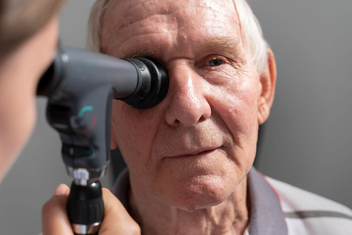
Diagnosis of Diabetic Retinopathy:
Due to the lack of symptoms in the initial stages, regular eye screening examinations are imperative for diabetic patients. Type 1 diabetic patients diagnosed before the age of 30 should commence screening five years after diagnosis. On the other hand, both type 2 diabetic patients and type 1 diabetic patients diagnosed after the age of 30 should initiate eye screening examinations at the time of diagnosis. These examinations involve pupil dilation using drops, allowing for a comprehensive retina examination with lenses. Additionally, an Optical Coherence Tomography (OCT) test, also known as eye tomography, is performed to assess the presence of diabetic macular edema. Early and regular screenings are vital for timely detection and effective management of diabetic retinopathy.
Treatment Approaches for Diabetic Retinopathy:
Diabetic retinopathy can be effectively treated through various methods.
Intraocular Drug Injections (Needle Treatment):
Current treatment methods for diabetic macular edema involve intraocular injections. Two types of injections are commonly used for this purpose. The first group includes anti-VEGF drugs such as Altuzan, Lucentis, and Eylea. While these drugs offer short-term effects, they temporarily alleviate abnormal vessel stretching in the proliferative phase. The second type involves Ozurdex implantation, a steroid-based treatment with a longer duration, although it does not shrink abnormal vessels.
Argon Laser Treatment
The primary treatment for abnormal new vessels in diabetic retinopathy is argon laser photocoagulation application. This treatment entails applying argon laser around the retina’s areas with reduced blood supply, contributing minimally to the visual field. The procedure typically spans 3-5 sessions to cover all targeted areas.
For a more in-depth understanding of argon laser treatment, refer to our article on “Argon Laser.”
Surgical Treatment (Vitrectomy Surgery):
Intense intraocular bleeding (vitreous hemorrhage) due to diabetic retinopathy may require up to one month for the bleeding to subside. If the bleeding persists beyond this period, vitrectomy surgery becomes necessary. Additionally, tractional retinal detachment, characterized by the development of membranes separating the retina from its position, may occur in diabetic retinopathy. In such cases, vitrectomy surgery is crucial.
For further insights into Vitreoretinal Surgery (vitrectomy surgery), consult our dedicated article on “Vitrectomy Surgery.”
Success Rate of Diabetic Retinopathy Surgery:
For diabetic retinopathy patients experiencing solely intraocular bleeding, the anatomical success rate reaches 100%, and vision levels can potentially improve to 1.0 (equivalent to 100%). However, when additional complications like diabetic macular edema and traction retinal detachment come into play, the potential for vision improvement becomes more limited.
Post-Surgery Considerations:
Following vitrectomy surgery, it is crucial for patients to maintain a balanced blood sugar level, in addition to other post-operative care guidelines.
For detailed insights into the recovery period and essential considerations after the surgery, refer to our comprehensive article on “Vitrectomy Surgery.”
Frequently Asked Questions About Diabetic Retinopathy
- Managing Diabetic Retinopathy During Pregnancy:
- Applying argon laser is safe if abnormal new vessels (neovascularization) develop due to diabetic retinopathy during pregnancy.
- For patients with diabetic macular edema during pregnancy, Ozurdex implantation is preferred over anti-VEGF agents.
- Link Between Diabetic Retinopathy and Cataract Disease:
- Cataracts tend to develop at an earlier age in diabetic patients.
- For patients with diabetic retinopathy, argon lasers should be completed if necessary before addressing cataracts.
- Cataract surgery may be more suitable after intraocular injection in patients with diabetic macular edema.
- Complications of Untreated Diabetic Retinopathy:
- Failure to treat diabetic retinopathy can lead to complete vision loss in the patient.
My Scientific Studies on Diabetic Retinopathy
Spectral Domain Optical Coherence Tomography Classification of Diabetic Macular Edema: A New Proposal To Clinical Practice (doi: 10.1007/s00417-020-04640-9): Published in the prestigious Germany-based journal “Graefe’s Archive For Clinical And Experimental Ophthalmology,” our study introduces a groundbreaking classification for diabetic macular edema. The comprehensive analysis, encompassing 408 eyes from 309 patients, revealed statistically significant findings. Notably, central foveal thickness exhibited a significant increase (P < 0.001), and there was a notable elevation in the rate of outer retinal damage (P < 0.001). This innovative classification promises to enhance our understanding and management of diabetic macular edema, contributing to advancements in ophthalmological research and patient care.
Henle Fiber Layer Thickness and Area Measurement in Type 2 Diabetes Mellitus With And Without Retinopathy Using A Modified Directional Optical Coherence Tomography Strategy (doi: 10.1097/IAE.0000000000003778): Our research, featured in the esteemed ‘Retina’ journal, a pinnacle in the realm of retinal diseases, achieved a breakthrough in measuring the area and thickness of the elusive Henle layer of the retina. Through a refined methodology, we overcame the challenges associated with imaging this intricate layer. The study’s findings demonstrated a statistically significant thinning of the Henle layer in the non-proliferative diabetic retinopathy group compared to both the diabetes group without diabetic retinopathy and the control group (all P < 0.05). This modification in measurement techniques opens new avenues for understanding retinal complexities, offering valuable insights for future research and clinical applications.

Causes and Treatment of Eye Floaters
Occasionally, individuals may notice floating objects in their field of vision. These can take various [...]
Read MoreMay

What is Vitreous? How is Vitreous Dysfunction Treated?
Vitreous, derived from the Latin term meaning “glassy,” is a clear gel found between the [...]
Read MoreMay

Vision Wellness Essentials: Simple and Effective Tips for Eye Health
Our eyes, vital and health-sensitive organs, demand special care to combat conditions such as eye [...]
Read MoreJan

Nourishing Your Eyes: A Guide to Beneficial Foods for Optimal Eye Health
Caring for our eyes extends beyond weight control; it encompasses exercise, abstinence from smoking and [...]
Read MoreJan

Understanding Glaucoma: Unveiling the Silent Threat to Vision
Glaucoma, also colloquially known as “black water,” refers to elevated eye pressure. This insidious eye [...]
Read MoreJan

What is Bietti Crystalline Dystrophy? Can Bietti Crystalline Dystrophy Be Treated?
Bietti Crystalline Dystrophy, identified by Italian physician Dr. Lucas Bietti in 1937, is a rare [...]
Read MoreJan

Understanding Color Blindness: Symptoms and Treatment
Color blindness is a vision condition where individuals struggle to distinguish specific colors or shades. [...]
Read MoreDec

What is Uveitis? What are Types, Symptoms, and Treatment Methods?
Uveitis, a severe eye condition characterized by inflammation, is also known as iridocyclitis. Accurate diagnosis [...]
Read MoreDec